
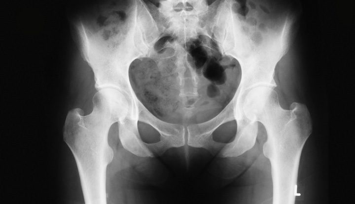
What Do You Notice? Understanding Pelvic X-Rays and Bone Abnormalities
Medical imaging, particularly X-rays, has long been one of the most important tools in modern healthcare. When a patient arrives at the hospital with severe pain in the hip or pelvis, an X-ray is usually one of the first steps in the diagnostic process. The image you see above raises an important question: What do you notice? Looking closely, you can see that the pelvic bones and hip joints show irregular changes, which may indicate fractures, degeneration, or other bone abnormalities.
In this article, we will explore the significance of pelvic X-rays, common causes of bone changes in this region, and why timely diagnosis is critical for recovery.

The Role of the Pelvis in the Human Body
The pelvis is a strong, ring-like structure of bones located at the base of the spine. It connects the upper body to the lower limbs, supporting weight when standing, walking, or running. It also protects vital organs such as the bladder, intestines, and reproductive organs. Because of its central role in movement and stability, any damage to the pelvis can have a profound impact on daily life.
What Doctors Look for in a Pelvic X-Ray
When radiologists and doctors examine a pelvic X-ray, they typically assess:
-
Bone Integrity – checking for fractures, cracks, or displacements.
-
Joint Spaces – looking at the hip joints to see if they are aligned properly.
-
Bone Density – identifying signs of osteoporosis or thinning of the bones.
-
Abnormal Growths – spotting tumors, cysts, or unusual bone formations.
-
Healing Patterns – evaluating previous injuries or surgeries to see if healing is taking place.
The irregularities visible in the provided image could be linked to several conditions, which we will now examine.
Common Conditions Detected in Pelvic X-Rays
1. Pelvic Fractures
Pelvic fractures can occur after high-impact trauma such as car accidents, falls, or sports injuries. In older adults with osteoporosis, even minor falls can result in serious pelvic breaks. Symptoms include:
-
Sudden, severe pelvic or hip pain
-
Difficulty standing or walking
-
Visible bruising and swelling
Pelvic fractures are considered serious because they may damage surrounding organs and blood vessels. Immediate medical care is required.
2. Osteoarthritis of the Hip
Another common finding in pelvic X-rays is osteoarthritis, a condition where the cartilage that cushions the hip joint wears away. Over time, this leads to:
-
Pain during movement
-
Stiffness in the hips
-
Decreased mobility
On an X-ray, osteoarthritis appears as narrowed joint spaces, irregular bone surfaces, and sometimes bone spurs.
3. Osteoporosis and Bone Weakness
With aging, bones naturally lose density, becoming more fragile. Osteoporosis often goes undetected until a fracture occurs. Pelvic X-rays may show thinning bone tissue and fractures that seem out of proportion to the trauma experienced. Preventive care, including calcium, vitamin D, and weight-bearing exercise, plays an important role in maintaining bone health.
4. Bone Tumors or Abnormal Growths
Although less common, pelvic X-rays can sometimes reveal unusual masses or irregular bone structures. These findings prompt further investigation with CT scans, MRIs, or biopsies to determine if the cause is benign (like cysts) or malignant.
Why Pelvic Fractures Are a Serious Concern
Unlike smaller bones in the body, the pelvis has a complex structure and is closely connected to major blood vessels and organs. A pelvic fracture can lead to complications such as:
-
Internal bleeding
-
Nerve injury
-
Damage to the bladder or intestines
Because of this, pelvic injuries are treated as emergencies in most medical centers. Depending on severity, treatment may involve bed rest, physical therapy, or surgery with plates and screws.
Recovery and Rehabilitation
Healing from pelvic injuries or hip joint issues can be a long process. Recovery often requires:
-
Pain Management – through medication and physical therapy.
-
Mobility Support – such as crutches, walkers, or wheelchairs in the early stages.
-
Rehabilitation Exercises – designed to strengthen muscles and restore flexibility.
-
Lifestyle Changes – maintaining a healthy weight, eating a nutrient-rich diet, and avoiding activities that put excessive stress on the hips.
In cases of severe hip arthritis, joint replacement surgery may be recommended, which can significantly improve quality of life.
Preventing Pelvic and Hip Problems
While some causes of pelvic injury are unavoidable (like accidents), there are steps individuals can take to protect their bones:
-
Exercise Regularly – activities like walking, swimming, and light weightlifting strengthen bones.
-
Eat Bone-Friendly Foods – including dairy products, leafy greens, and fortified grains.
-
Avoid Smoking and Excessive Alcohol – both can weaken bone structure.
-
Monitor Bone Density – especially for older adults or those with a family history of osteoporosis.
Conclusion
Looking at the pelvic X-ray above, it is clear that the bones show signs of irregularity, possibly due to fractures, arthritis, or bone degeneration. These findings highlight how vital X-rays are in detecting hidden problems and guiding doctors toward the right treatment.
The pelvis may seem like just another part of the skeletal system, but it plays a central role in movement, balance, and organ protection. Whether the issue is a traumatic fracture or age-related degeneration, early diagnosis and treatment make all the difference.
So, the next time you come across a medical image that asks, “What do you notice?”, remember that each detail in an X-ray tells a story about the body’s health. By learning to recognize the signs, patients can better understand their conditions and work with doctors toward recovery.